
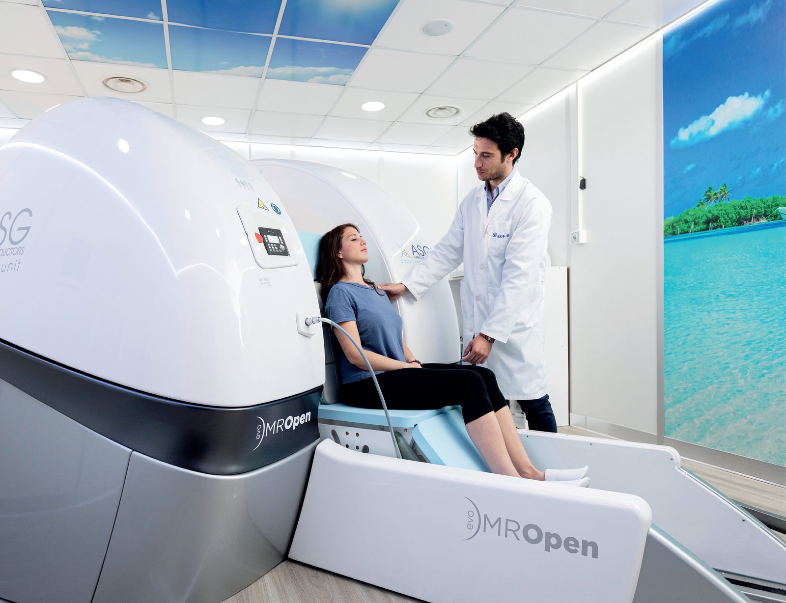
MROpen Evo — the open, helium-free MRI
MROpen Evo is the 0.5 tesla magnetic resonance scanner built by the Italian ASG Superconductors (Paramed MRI Unit). Two things set it apart from conventional tunnel-type MRI systems: the open, U-shaped magnet, in which the patient is not enclosed in a bore, and the magnesium diboride (MgB₂) superconductor, which operates without helium.
Belux Csoport distributes the system in Hungary — from installation and commissioning through operator training to ongoing support.
Important
In MROpen Evo the patient walks into the scanner and can be imaged sitting, leaning back, lying down or standing. There is no physical barrier between the patient and the surrounding room: they can see around them at all times and can even watch TV during the examination. This fundamentally changes what is possible with claustrophobic, anxious, paediatric, elderly and larger patients.
Upright and multi-position imaging
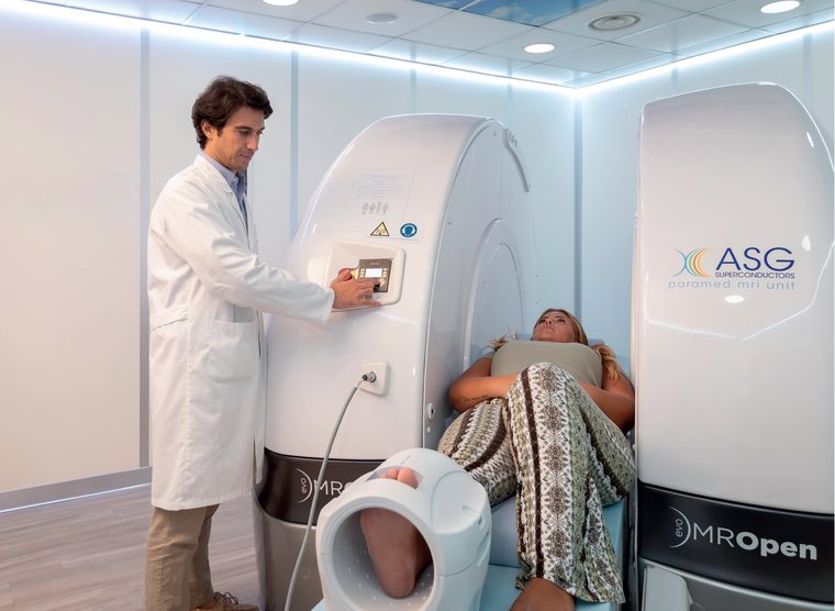
A conventional MRI can only image the patient lying down — that is, in exactly the position in which the symptoms usually do not occur. MROpen Evo enables symptom-driven, positional imaging:
- Weight-bearing imaging — the spine and joints can be imaged under real body-weight load, in the standing position.
- Functional studies — the spine and joints can be imaged in flexion and extension.
- Positional imaging — the patient is scanned in the position of maximum symptom: standing, sitting, bending or lying down.
The scanner works as a stand-alone system and as a complement to a conventional MRI, taking over the examinations a closed bore cannot perform, or can perform only with difficulty.
Anatomies and patient positions
| Area | Examinations | Patient positions |
|---|---|---|
| Cervical spine | routine, flexion/extension, soft tissue neck, carotid MRA | supine, 20°, 40°, sitting |
| Thoracic and lumbar spine | routine, flexion/extension, weight bearing | supine, 20°, 40°, 60°, sitting, standing |
| Brain | brain, internal auditory canal (IAC), temporomandibular joint (TMJ), pituitary gland, orbits, MRA | supine, 20°, 40°, sitting |
| Knee | routine and weight bearing | supine, 20°, 40°, 60°, sitting, standing, bent knee |
| Extremities | hand, wrist, elbow, ankle, foot, shoulder | supine, 40°, sitting, standing |
| Hip | routine and weight bearing | supine, standing, standing with hip rotation |
| Pelvis | routine | sitting, supine |
MgB₂ — superconduction without helium
The MROpen Evo magnet is built from magnesium diboride (MgB₂) high-temperature superconductor with helium-free cooling. This has direct operational consequences:
- No helium refills and no venting line — magnet installation and day-to-day operation are considerably simpler than with conventional solutions.
- Quench-free, stable operation — the magnet is fully controllable, including switching on and off, with simplified start-up and maintenance.
- Lower power consumption and a much reduced use of rare-earth elements.
Receiving coils
| Coil | Channels | Clinical application |
|---|---|---|
| Whole Spine | 8 | whole spine |
| Body Spine | 4 | L-spine, T-spine, pelvis, bilateral hips, sacroiliac joints |
| Spine Flat | 4 | upright/sitting L-spine and T-spine, sacroiliac joints |
| Cervical Spine | 2 | C-spine |
| Head Neck | 3 | brain, MRA |
| Brain | 2 | head, brain |
| Knee | 2 | knee, ankle, foot |
| Wrist-Hand | 2 | hand, wrist, elbow |
| Shoulder | 2 | shoulder |
| MP-Loop | 1 | shoulder, dynamic studies, multipurpose |
| MP-Flat | 1 | upright/sitting hip, pelvic floor, multipurpose |
| Flex Small / Large | 1 | upright/sitting pelvis, bilateral hips |
Pulsed sequences
- Spin echo — Spin Echo, Gradient Echo, Multi Echo, PD-T2, Gradient Echo Dual Echo (HiCon)
- Fast spin echo — FSE T2, FSE T1, FSE PD, Fast Inversion Recovery (FIR PD, FIR T2), Fast FLAIR, Metal T1/T2, HASTE, diffusion weighted imaging (DWI–HASTE)
- Gradient echo steady state — GBASS 3D/2D, GE 3D, GE T1 3D, EMIT 3D, STSS 3D
- MRA — Angio 2D, Angio 3D
- Fat suppression — Fat Water Separation (FWS), GE-STIR, SE-STIR
Technical specification
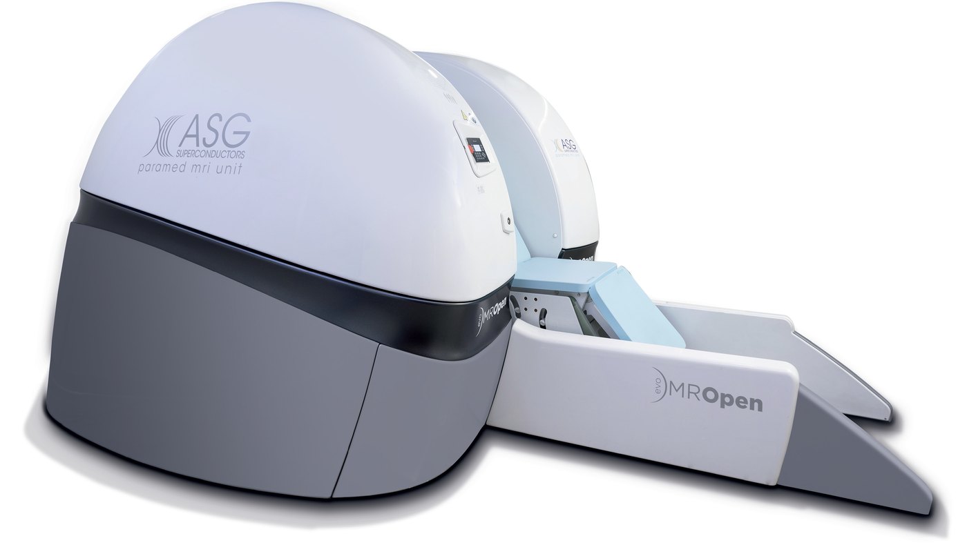
| Parameter | Value |
|---|---|
| Field strength | 0.5 T |
| Magnet type | superconductive, cryogen-free, based on MgB₂ (magnesium diboride) technology |
| Lateral gap | 56 cm |
| Digital spectrometer | I-box |
| Receiving channels | 8 |
| Gradients max. intensity | 20 mT/m |
| Rise time | 0.6 ms (0–20 mT/m) |
| Slew rate | 33 mT/m/ms |
| Coil technology | multi array cross tuned |
| Max. patient weight | 200 kg |
| Patient position | laying, sitting, standing |
| Graphic user interface | MR-GUI Pro |
Siting requirements
| Parameter | Value |
|---|---|
| Total system weight | 28,000 kg |
| Magnet room, standard dimensions | 6 × 5 m |
| Technical room, standard dimensions | 3 × 4 m |
| Electronic cabinets | 3 × 250 kg |
| RF shielding | required |
| Power requirement | 400 V 3N ~50 Hz / 480 V 3N ~50/60 Hz |
| Power consumption | 35 kVA |
Note
Room and power requirements are always planned against the actual site. The figures above are the manufacturer's standard dimensions; we provide detailed plans after a site survey.
Deployment and support
We carry out the site survey, installation, commissioning, operator training and ongoing technical support. The scanner fits into the existing radiology workflow: images are sent in DICOM format to the PACS, and reporting happens as usual, supported by speech recognition if required.
Ask for a quote or a demonstration — or visit the manufacturer's site at mropenevo.com.